This article has been reviewed according to Science X's editorial process and policies. Editors have highlighted the following attributes while ensuring the content's credibility:
fact-checked
proofread
Researchers highlight advancements in biomedical research with enzyme-activated fluorescent probes

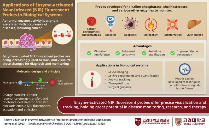
Enzymes, essential for normal cellular and physiological functions, are implicated in various diseases like cancer and diabetes due to their abnormal activity. Therefore, tracking enzyme activity is a valuable strategy for the diagnosis and monitoring of diseases. Conventional imaging techniques are limited by the need for contrast agents, low sensitivity, and spatio-temporal resolution.
To overcome these limitations, researchers are increasingly investigating fluorescent probes for non-invasive and real-time visualization of enzyme dynamics and corresponding disease status.
In a new review article, researchers from Korea have summarized the latest advancements in the development of enzyme-activated near-infrared (NIR) fluorescent probes and their diverse applications in biomedical research and medicine. Providing further insight into their article, the lead authors,
Professor Jun-Seok Lee from Korea University College of Medicine and Professor Juyoung Yoon from Ewha Womans University in Korea, explained, "While conventional biomarker examination relies on the comparative expression level of a target enzyme, it does not reflect enzymatic activity. Enzyme-activated fluorescent probes can help monitor the dynamics of enzyme activity in vitro and in vivo."
The review article was published in Trends in Analytical Chemistry.
Enzyme-activated fluorescent probes primarily consist of three components: a fluorophore that emits fluorescence upon activation, a linker, and an enzyme recognition unit. When the probe encounters the target enzyme, the resulting charge or energy transfer activates the NIR-fluorophore, emitting detectable fluorescence.
The authors describe various design strategies for these adaptable fluorescent probes, with versatile applications in studies targeting enzymes involved in metabolic processes, neurotransmission, cell growth, cell death, and other key processes.
NIR-fluorescent probes are widely used in biomedical imaging to visualize cells and tissues, and they provide highly sensitive and real-time measurements of enzyme activity in cells as well as animal disease models.
Their selectivity allows for the detection of aberrant enzymes specific to certain tumors or diseases, aiding early and differential diagnosis. Additionally, they are used to outline tumor margins or specific tissues, guide surgical resection, and hold promise in assessing therapeutic responses to enzyme-targeting therapies. Furthermore, their applications extend to environmental sensing, food safety, water, and air analysis.
Enzyme-activated fluorescent probes offer high specificity and sensitivity, excellent biocompatibility, ease of use, and tunable properties, making them valuable assets in biomedical research and health care. Further studies can help in the designing of multi-target fluorescent probes capable of distinguishing between different cell types and aiding in clinical research, diagnostics, disease monitoring, and treatments. Overall, these probes hold significant potential to revolutionize health care.
This review enhances our understanding of fluorescent probes and lays the foundation for future research that can expand their applications. The authors conclude by saying, "Abnormal enzyme activity is a hallmark of several diseases. NIR fluorescent probes can be used as molecular tools for visualization and quantification of such biomarkers. While significant advances have been made in their development, additional studies are needed to widen their bioapplications."
More information: Hyunsun Jeong et al, Recent advances in enzyme-activated NIR fluorescent probes for biological applications, Trends in Analytical Chemistry (2023). DOI: 10.1016/j.trac.2023.117335
Provided by Korea University College of Medicine


