Under attack: Researchers shed light on how Lyme disease infects body

An estimated 476,000 Americans are infected each year with Lyme disease, a condition causing a wide range of symptoms that include fever, rash, and joint pain, as well as effects on the central nervous system and heart. Though it's common knowledge that Borrelia burgdorferi—the bacteria that causes the disease—enters the body through the bite of an infected deer tick, how the bacteria manages to migrate from that bite into a person's bloodstream has not been clearly understood.
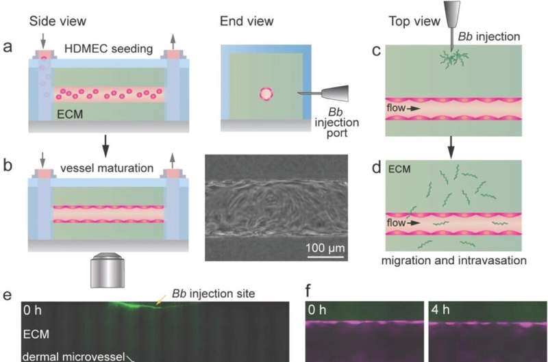
Johns Hopkins engineers may have found the answer. Using a custom designed three-dimensional tissue-engineered model, they learned that B. burgdorferi uses tenacious trial-and-error movements to find and slip through tiny openings called junctions in the lining of blood vessels near the original bite site. This allows them to hitch a ride on the bloodstream throughout the body, potentially infecting other tissue and organs. Their results appear today in Advanced Science.
"Our observations showed that if the bacteria did not find one of these junctions on the first try, they continued searching until one was found," said team leader Peter Searson, professor in the Whiting School of Engineering's Department of Materials Science and Engineering and core researcher at Johns Hopkins Institute for NanoBioTechnology. "The bacteria spend an hour or two using this behavior to find their way into the blood vessels, but once there, they are in circulation in a matter of seconds."
The team injected its three-dimensional model, which simulates a human blood vessel and its surrounding dermal tissue, with the bacteria, simulating a tick bite, and used a high-resolution optical imaging technique to observe its movements. They observed that though the tissue at the original bite site was an obstacle for the spiral-shaped bacteria to surmount, little effort was needed for them to penetrate the junctions and enter the bloodstream.
"We've previously created tissue-engineered vascular models of other tissues, so we applied what we learned to create a model of dermal tissue to study the behavior and mechanisms of dissemination of vector-borne pathogens," said Searson.
Lyme disease is prevalent in North America, Europe, and Asia, and though antibiotic treatments are effective, some patients experience symptoms that can persist for months, and, in some cases, years. The Hopkins team says that understanding how B. burgdorferi spreads throughout the body could help inform treatments to prevent bacteria from the initial bite entering other tissues and organs.
"We also believe that the kind of human tissue-engineered model we created can be broadly applied to visualize the details of dynamic processes associated with other vector-borne diseases and not just Lyme disease," Searson said.
More information: Zhaobin Guo et al, Visualization of the Dynamics of Invasion and Intravasation of the Bacterium That Causes Lyme Disease in a Tissue Engineered Dermal Microvessel Model, Advanced Science (2022). DOI: 10.1002/advs.202204395
Journal information: Advanced Science
Provided by Johns Hopkins University


